
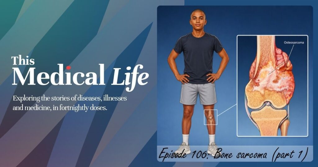
Bone sarcomas are rare, accounting for 0.2% of all cancer diagnoses, with around 200 cases diagnosed in Australia each year. The three main types differ by age and site: osteosarcoma (around 30 to 35% of cases, often in the arms, legs and pelvis, with peaks in adolescence and again in the seventies and eighties), chondrosarcoma (around 30% of cases, typically in adults over 40), and Ewing sarcoma (around 15% of cases, most common in children).
Professor Fiona Maclean, an anatomical pathologist, describes bone sarcoma diagnosis as a pattern-recognition challenge made harder by rarity, noting that tumour type and stage are the two most useful pieces of information for a GP reading a pathology report.
A/Professor Richard Boyle, an orthopaedic oncological surgeon, outlines the red flags, including unexplained deep bone pain and night pain, and the diagnostic pathway from X-ray through biopsy to multidisciplinary review.
This is the story of Bone Sarcoma Part 1.
Useful links:
Australia and New Zealand Sarcoma Association: https://sarcoma.org.au/
Find a sarcoma specialist: https://sarcoma.org.au/pages/about-sarcoma/find-a-sarcoma-specialist
Sarcoma guidelines: https://sarcoma.org.au/pages/sarcoma-guidelines
Seeking a second opinion: https://sarcoma.org.au/pages/about-sarcoma/seeking-second-opinion
Useful patient information: https://sarcoma.org.au/pages/about-sarcoma/patient-and-carer-sarcoma-information
Our Special Guests:
A/Professor Richard Boyle who is a Sydney-based orthopaedic oncological surgeon and head of the NSW Sarcoma unit.
Professor Fiona Maclean who is an anatomical pathologist and professor from Macquarie University.
Listen:
This Medical Life podcast is available on all podcasting services, Apple Podcasts, YouTube, and Spotify. Members of the RACGP are able to log the hours as CPD (Education) that they listen to the episodes under self record via Quick log. We invite you to help support us on our donation page.
Automated Transcript:
This transcript was generated automatically by Descript. I will contain errors and spelling mistakes, but is offered as a guide and resource to help AI and search tools discover the content on behalf of GPs, medical students, other allied health professionals, and the general public.
TML S07E106
Steve Davis: [00:00:00] Welcome to This Medical Life podcast. These are the stories of medicine with Steve Davis and Dr. Travis Brown. This is the story of bone sarcomas, part one
Dr. Travis Brown, we’ve just spent two episodes on sarcoma, soft tissue sarcoma. We’re changing our scenery
Dr Travis Brown: We are. We’re still staying on the s- focus of sarcoma. and we’re going to talk about bone sarcoma. Now, bone sarcomas are rare. so this fits- Hang on,
Steve Davis: hang on. Even soft tissue sarcomas are rare is what you said
Dr Travis Brown: And bone are even rarer, if, if that’s a word.
and it’s, it’s one of those ones [00:01:00] that you sit there and just go, look, there’s only, like, a few hundred cases, around 200 cases diagnosed in Australia a year. If you look at the United States, a bit over 4,000 a year. So these are, these are rare. and, they account for 0.2% of all cancer diagnoses. And so this was one of those, one of those things that In pathology, you, you almost, well, at least I did, you study by, by numbers.
Like, you look at it and you just go, when you’re looking at a biopsy down the microscope, whether it be, whether slides or cases or y- or reporting, in the back of your mind you’re going, “Well, how common is the diagnosis that I’m about to present?” Because if this is a super, super rare, condition, is it really rare or am I just seeing something that’s not there and, you know, it could be a much more common presentation, just an odd one.
And so this is genuinely, when you look at bone, [00:02:00] you actually find that the, the, the likelihood of it being a metastasis is far higher than it being a primary m- primary bone malignancy. And so this is because, you know, commonly there are a few cancers that go to bone, so breast, lung, prostate. It’s classic for them to actually turn up as a metastasis in the bone.
and so this is why when we get a history, you actually say, “Have they got a history of malignancy” when you’re looking at these bone lesions. And but when you come to, like, bone sarcomas, the, the amazing thing is there’s not much historically, i- in the records, in medical literature, and this is because they were, first of all- They wouldn’t have been, yeah
they were rare. Yeah, they, they were rare. But also it’s one of those things that, it’s m- kind of a modern diagnosis because of our investigations that we can do. So, when X-ray came along at, you know, end of the 19th century, start of the 20th century, then you could actually start to look at bones and [00:03:00] see bones as opposed to-
Steve Davis: So is this one of those rare examples in this medical life where you’re not taking us back to ancient Egypt-
where the doctors first discovered it?
Dr Travis Brown: I, I, I’m not, and that’s the thing. If you look at the archeological history, because bones actually are preserved quite well, there’s, there’s a few scattered instances. Like, there’s a, there’s a y- 240 million-year turtle that has believed to have had a sarcoma. And- Wow
there’s a, there’s a, actually there’s a really interesting case that’s come out of, Swartkrans Cave, which is in near Johannesburg in, South Africa. Now, this is actually a cave system that pretty much it’s either, it was either a natural trap or a, a, an area that collected bones, and they’ve found, the, i- it’s called SK s- not, sorry, SK7923.
Now this is a metatarsal of a, human ancestor, and they believe that this fifth metatarsal has sarcoma in it. And it’s, [00:04:00] they’ve estimated about 1.7 million years ago. And, and this sort of feeds into that area where it talks about, oh, is cancer just a recent diagnosis or, th- this suggests- Yeah … as we’d expect, no, it’s not.
Cancer’s been with us for a long time. and- Again, it’s one of those things that difficult to find. So, of, of everything when we’re talking about bone sarcoma, we’re talking about three diagnoses. So there is actually osteosarcoma. it, it accounts for about thirty to thirty-five percent of cases. It tends to– It can occur in any bone, but it tends to be in the arms and legs and pelvis.
there’s a bimodal, age presentation, so there’s peaks in, the second decade, of life, so children, adolescents, and then patients between the ages of seventy and eighty. And so we– when we look at chondrosarcoma, this accounts for about thirty [00:05:00] percent of bone sarcomas. this affects upper arms, legs, pelvis, ribs, and the scapula, so the shoulder blade.
but this tends to happen for people over the age of forty. And so the, the last one, which we’ll actually discuss a little bit more at the start of next episode as well, is Ewing sarcoma. Now, this accounts for fifteen percent. This can occur in, in the pelvis, in the legs, ribs, spine, upper arms. Can occur at any age, but this is the one that tends to affect children more than anyone else.
Steve Davis: All those different modes of where they peak, plus they’re being rare, I mean, hats off to medical science to even discover these things in the first place.
Dr Travis Brown: And so these days, we’re actually really good at managing all of these conditions. we have, first of all, because they are rare, we have subspecialties, and specialists dealing with this.
we have sarcoma, multidisciplinary meetings. This will happen in countries, where they actually talk. And again, it’s [00:06:00] actually looking at diagnosis and management, treatment, what is going to be best for these patients. And so this is why we’re discussing, first of all, bone sarcoma in part one. and we will see.
We’ll talk to the experts and see what we can learn for that for, for, for doctors and for patients who, encounter this.
Steve Davis: Associate Professor Richard Boyle is a Sydney-based orthopedic oncologist and surgeon dealing with management of benign and malignant bone and soft tissue tumors. He works at North Shore Private and RPA, as well as the Children’s Hospital Network, and is head of the New South Wales Sarcoma Unit.
Associate Professor Boyle is a fellow of the Australian Orthopedic Association and is current Sarcoma Sub-Specialty president. Dr. Boyle is also medical [00:07:00] director of the New South Wales Bone Bank, associate professor at University of Notre Dame, and senior lecturer at the University of Sydney. And Richard’s a returning guest in our series about sarcomas with This Medical Life podcast.
Richard, welcome back.
A/Professor Richard Boyle: Thanks very much, and thanks for having me back. I obviously didn’t make too many mistakes the first time.
Steve Davis: No, yeah, you’re in danger of being invited numerous times in the future.
A/Professor Richard Boyle: Right. Okay.
Steve Davis: Richard, how do you approach a patient who’s been diagnosed with bone sarcoma?
A/Professor Richard Boyle: Excellent question, and we can probably just answer that over the next half an hour.
the … Perhaps if I take a step back in that often they … A patient won’t necessarily turn up and say, “My GP’s referred me with an osteosarcoma.” There’s, there’s a bit of a process that gets to that diagnosis. and in, in sort of, in very brief terms [00:08:00] A GP will have seen them. There may well have been a lump or some pain and so forth that they’ve noticed that’s a firm lump that’s a bit concerning.
Perhaps they’ve returned a few times after a simple injury, and it’s just not getting better, and they’ve had pain at night. And an X-ray is done, and on the X-ray there’s some concern, and maybe on the report it says something like a sarcoma should be considered. Often that’s when the referral comes through to me, and then that will, stimulate a chain of further investigations, that may well involve MRI scanning, of the lesion itself, and then also, what we would call a systemic staging scan, such as a PET scan.
back in the day it would have been, bone scanning, but over the last realistically 20 years, PET scanning has become the modality of choice. and I’ll go into that a little bit later on. And then a biopsy, and the biopsy is done very, very [00:09:00] carefully, and at a center generally that will have experience in bone pathology, but also at the site usually where further treatment will occur, be that chemotherapy and/or radiotherapy and/or, surgery.
Radiotherapy not so much for osteosarcoma. and, the entire case is discussed at what’s called an MDT, the multidisciplinary team meeting, where all of the imaging and pathology is presented, and then, the medical oncologist, radiation oncologist, surgeons, so forth are all in the room, as well as the allied health team.
And a final diagnosis is made, and then a management plan is put into place. So th- that’s the very extensive process that goes into the point of getting to saying to someone, “Yes, this is an [00:10:00] osteosarcoma.”
Dr Travis Brown: In the soft tissue episode, we discussed whoops lesions, so doctors willing to give something a go with regards to soft tissue that maybe would be better if they had left it to, to experts i- in this area.
I wouldn’t imagine … Is there many doctors who would actually give a go of a biopsy on a bone lesion at all, or even unexpectedly biopsy it?
A/Professor Richard Boyle: Yeah. So that certainly usually falls into the realm of other surgeons, not so much in general practice or, you know, community sort of centers. But, it does occur unfortunately.
In that, as you might have sort of picked up on there, the whole process to get to the diagnosis is very complicated That’s because sometimes it can be subtle, or sometimes it can be masquerading as a different condition such as an infection in bone. and one of the challenges in bone sarcoma is that one of the highest peaks of incidence is in, teenage years or adolescent young [00:11:00] adult years.
And that’s also the peak when you will have other conditions such as osteomyelitis. And an infection in bone can look very similar to what a sarcoma may well look, on X-ray and MRI. and it may well be that a surgeon takes them to theater expecting to find an infection, and it’s actually a tumor. and yes, unfortunately that does occur.
but, it can be difficult, it can be difficult to distinguish.
Dr Travis Brown: Now we’re lumping these all together, so I … We can, we can s- tease them out a little bit, but is there any common or classic presentations of, of osteosarcoma or chondrosarcoma or Ewing sarcoma?
A/Professor Richard Boyle: Just going back to that point there about the epidemiology in that chondrosarcoma is generally a presentation of, adult life, whereas Ewings and osteosarcoma is far more common in the younger age bracket.
and so in that [00:12:00] age bracket, so now if we just focus on osteosarcoma Ewings, it’s sometimes, many months of just an unexplained pain, a deep pain. very early on I think I had a patient say to me, “It feels like a toothache in the bone.” and I’ve now used that ever since then because it’s sort of a good descriptor in that people can relate to that.
pain at night rather than activity-based pain. And sometimes, and perhaps more with Ewings than osteosarcoma, they can have some of those more systemic features of weight loss and lethargy and so forth, but, many occasions they don’t. It’s just purely the symptoms at the local site.
Dr Travis Brown: Are there any risk factors for developing sarcomas, bone sarcomas?
A/Professor Richard Boyle: There are some familial cancer syndromes, that you may have heard of, you know, associated with retinoblastoma gene, [00:13:00] Li-Fraumeni syndrome. But in most cases, we don’t know what the cause is. so in elderly life, yes, there are some other associations with other conditions, which is very, very rare, but things like, prior exposure to, radiotherapy, is definitely a, a risk.
perhaps more leaning into an example of angiosarcoma post-radiotherapy for breast cancer. but the bony sarcomas, yeah, things like Paget’s disease, bone infarcts and so forth, but exceedingly rare.
Dr Travis Brown: Are there any presenting symptoms that are, are red flags for you that says, “Oh, this needs to be dealt with immediately”?
is there anything?
A/Professor Richard Boyle: Well, pathological fracture. If someone presents with a fracture through a lesion, then obviously that’s very problematic. Yeah, severe pain, in a, in a child, the, that does need to be dealt with [00:14:00] straight away. Yeah, I mean, it’s, there’s not really any bone sarcoma that we would say, “Oh, let’s just watch this.
Come back in a few weeks time.” We’ll get onto it straight away, and, you know, best case scenario is diagnose it as an infection or diagnose it as a benign lesion. You know, eosinophilic granuloma is another one that can look quite aggressive, but, Yeah, we, we, it, it’s very unlikely that we would sit and wait on a sarcoma or something that is suspicious for it.
Steve Davis: It’s not very common for us to talk about amputation on This Medical Life, but in the re- the interviews we’re recording for this episode, it has come up. Yes. What determines whether a patient will have limb-sparing surgery or amputation?
A/Professor Richard Boyle: So obviously, that’s a, a very complex decision process, but, as a, as a sort of simplified answer, if you’re going to save a limb, it needs to be a functional limb.
And so if there… Or actually, [00:15:00] just taking a step back from that, it needs to be oncologically resectable. So in other words, I need to make sure that I can actually get all of the tumor out at surgery, and if I cannot do that, then it needs to be an amputation. but then also, let’s say technically we can get around the tumor, for want of a better term, and I’m taking out all of the nerves and blood vessels of a leg, then there’s no point.
Or if it means that you have to reconstruct most of the skeletal elements and remove, a major functional part of the leg, such as the quadriceps or the extensor mechanism of the knee, then again, you’re actually better off with an amputation. And it’s a bit confronting, and sometimes hard to explain, but the younger the child, often the better they cope with an amputation because then that becomes their normal [00:16:00] life.
it’s harder, the older the patient is.
Dr Travis Brown: So when we’re looking at, treatment, when you’re looking surgically, if you were to treat a child who had Ewing sarcoma and osteosarcoma in exactly the same spot, is there any difference surgically, or it’s just the question can I remove it all?
A/Professor Richard Boyle: So Ewing sarcoma actually responds relatively well to radiotherapy.
and there’s a lot of research now around, for example, pelvic Ewing sarcoma that shows better outcomes in combination with radiotherapy. So often that’s an example where we will very strongly consider radiotherapy and surgery and chemotherapy, whereas osteosarcoma doesn’t respond so well to radiotherapy.
so in that situation we would, You very carefully look at and lean [00:17:00] much more towards, surgery. I should say both Ewing’s and osteosarcoma will always have what’s called induction chemotherapy, so in other words, chemotherapy first, and then whatever is to be the form of local treatment, be it radiotherapy, surgery, combination.
the other consideration is how much of the bone has been, eaten away or destroyed by the tumor. and osteosarcoma Just by the, the fact that it is a bone-forming tumor, often the bone is actually, reusable, and we use a technique, that was really pioneered in Australia by, my mentor and senior colleague, Paul Stallys, who’s now retired.
But Paul brought this technique back to Australia where the segment of bone that is affected by the tumor [00:18:00] is irradiated, so resected and then irradiated, and then that is used back in the patient. So it’s sterilized and so forth, so it’s safe to use. and almost always we will all add into that, what’s called a vascularized fibular graft, that brings blood supply back to that bone, over time.
So that’s a technique that is more commonly used, and we do that as much as we can here in Sydney because that’s termed a biological reconstruction. sorry, I’ve gone off tangent a little bit there. I hope that answered the qu- the question that you asked.
Dr Travis Brown: No, no, that’s great. just in, just for a broad stroke, when patients present, is it
How common is it for them to present with advanced disease?
A/Professor Richard Boyle: Yeah, we do see that. and that’s disappointing, and it also confers a poorer prognosis. look, that … It- it’s very variable. We will go through a, a, [00:19:00] a, a series of, you know, patients, a couple of patients in a row just recently who presented with advanced disease.
but I don’t think I’ve got a hard and fast rule or answer to that in that, it’s, it’s almost impossible to predict or prevent that from occurring in that patients don’t know, and especially, you know, kids and teenagers don’t necessarily know that they’ve got a tumor and would not expect that they’ve got a tumor either.
Dr Travis Brown: Are there any useful blood tests i- in this area?
A/Professor Richard Boyle: No, not yet. Not yet. and that goes back to, you know, the explanation of sarcoma being a mesenchymal-based tumor in that bones, muscles, fat tissues, so forth don’t produce anything. Whereas carcinomas such as prostate produce things. So prostate you can measure PSA.
So, and there aren’t any [00:20:00] tumor cell markers like there are in many of the carcinomas. So no, there’s not, unfortunately not a blood test yet. but that’s a whole area of research called liquid biopsy, where in the future I’m sure this will come in where we’ll be able to hunt down foreign DNA or, you know, from pieces of, DNA, that will be an indication that there is, sarcoma or recurrence of sarcoma, you know.
And, and then scans like PET scans, may well become a thing of the past, or at least less commonly used as surveillance scans.
Steve Davis: Richard, just a moment ago, Travis asked you about, patients presenting with advanced, forms of the disease, but it prompted another question. What sort of questions do patients ask you when they’re presented or suspected, if you like, of having a bone sarcoma?
A/Professor Richard Boyle: Yeah. So obviously the question that they want [00:21:00] answered is, am I going to live? And often that’s danced around with all sorts of other things. You know, I play soccer, will I be able to play soccer next season? You know, those sorts of things. but, usually it will be, am I going to lose my leg? Am I gonna need chemotherapy?
Am I going to be sick? Will I get sick on the chemotherapy? Unfortunately, the answer is yes. but as a general rule, upfront I try to remain as hopeful and positive as possible. and say that, you know, our intention is to perform, as you’ve termed there, limb salvage surgery, and to get through it and, and to beat it.
And, and look, the percentages are good but not great. as a very, very rough sort of number, the overarching success rate is perhaps 70 to 80%. [00:22:00] but if you flip that around, it means, you know, one in five of these kids doesn’t survive. and that’s where we need more research, and we need more funding for research, and I think we can do better.
Dr Travis Brown: What’s the best measure of prognosis you use when you’re looking at osteosarcomas, chondrosarcomas, or Ewing sarcoma?
A/Professor Richard Boyle: Your previous question is, is probably the number one, how advanced it is. so if they present with metastatic disease, then that’s a problem. Response to chemotherapy is the next most important thing realistically.
and I’ll often have a joke with colleagues or even some of the patients, depending on if it’s the correct scenario to say, even though surgeons think they’re the most important people in the world, I have to admit it’s actually the medical oncologists with the chemotherapy, who are the ones that win the day.
if the tumor does not respond to chemotherapy, then the [00:23:00] surgery is not the thing that’s gonna save their life, unfortunately, or very, very unlikely to, unless it’s localized and you’ve been able to get around it. Sometimes we will come in early if it looks like the chemotherapy isn’t working, and we’ll operate, and if it hasn’t spread at that point, then we may well be able to save them.
But, yeah, there’s a, there’s a general sort of, you know, overarching comment, the prognostic factors, that are most important. yes, advanced disease, so metastatic disease or not, response to chemotherapy. tumor grade comes into it when you’re talking about chondrosarcoma. less so with osteosar and Ewings.
There are some rarer subtypes of osteosarcoma that portend a better prognosis, yes. and perhaps tumor site as well, in that pelvic tumors often will be larger, [00:24:00] and more difficult to resect just because of their anatomical location. and similarly so, you know, spine and, you know, hip and neck-based sarcomas as well.
Steve Davis: Do you have any final thoughts, Richard, for, doctors or even patients about bone sarcomas?
A/Professor Richard Boyle: The one thing to say is think about it or just consider it. In those scenarios where there’s the red flags of a returning patient that you’re just not sure about, it’s very straightforward to just order an X-ray, or even go a step beyond that and ask for help, from either your local orthopedic surgeon, perhaps all of the sarcoma centers around Australia, are very accessible.
And, I’m in the fortunate position of having known all of the sarcoma [00:25:00] tumor, sarcoma surgeons and orthopedic oncologists around the country, and they’re all fantastic people and would definitely much rather take a phone call and give reassurance than take on a patient with advanced disease that perhaps could have been seen earlier.
Steve Davis: Associate Professor Richard Boyle, thank you for joining us on This Medical Life.
A/Professor Richard Boyle: Happy to. Happy to come back if you’d really, really like me to.
Steve Davis: Professor Fiona McLean is a clinical professor in the Department of Clinical Medicine, Faculty of Medicine and Health Sciences, Macquarie University. She’s authored more than 100 peer-reviewed papers, as well as two chapters of Sternberg’s Diagnostic Surgical Pathology, and one chapter of Histology for Pathologists.
[00:26:00] Professor McLean is a previous president of the Australasian Division of the International Academy of Pathology, with interests in urogenital and musculoskeletal pathology. Fiona’s a regular guest here on this Medical Life podcast. Fiona, welcome back.
Professor Fiona Maclean: Well, thank you for having me again. I enjoy being referred to as a regular guest, so, that’s very nice.
Steve Davis: May it continue. Thank you. so we’re talking of regular, we’re continuing our topic of sarcoma, but this time bony sarcoma. How difficult is it to diagnose a bony sarcoma?
Professor Fiona Maclean: For people who had listened to the soft tissue sarcoma, podcast that we had earlier, I talked about, you know, it being a rare tumor.
So it, it is hard to identify something you’ve never seen before or you’ve only seen irregularly because, you know, anatomical pathology is basically pattern recognition, so if you haven’t seen a pattern, it’s very hard to see, yeah, to be able to identify it. bone sarco- [00:27:00] sarcomas are even more rare than soft tissue sarcomas, so they’re, you know, only 0.2% of all neoplasms.
And they’re about one-tenth as common as what I was referring to as rare last time, as the soft tissue sarcoma. So we’re even in a, a rarer category. So, experience, I think, is the issue with, with these tumors. You know, a lot of people don’t, receive bone specimens, you know, in their practice, and those that do, you know, may be seeing, mainly benign conditions.
So being able to identify a sarcoma for what it is is quite difficult. I was doing some AI work recently, and we, we really required about 100 examples of something to train the AI, like 100 examples of each entity, and there’s many bone sarcomas that I would report in my entire career that I’m not gonna see 100 of.
So you know, their, their rarity does make it difficult. You know, thinking we can’t train AI on that at this stage, we haven’t got to that stage of being able to, you know, get the AI to, to train on smaller numbers. [00:28:00] so I’m expected though to be able to identify it, you know. There’s a bit, ironic I think.
Steve Davis: Well, human intelligence is the superior version.
Professor Fiona Maclean: Of course.
Dr Travis Brown: Of- So we’re mainly talking about then osteosarcoma, chondrosarcoma, and Ewing’s sarcoma. How difficult is it to distinguish these, these entities?
Professor Fiona Maclean: Yeah, so last time I was talking about, you know, we really look at the morphology, the shape of the cells, to try and work out what sort of cell type the cell has come from.
and look, Ewing sarcoma is a bit different because it’s what we call a small, round, blue cell tumor, and so it, it, it’s, just that. Like, it’s very descriptive. We like to be descriptive in pathology. so it is actually just that. It is a small, round, blue cell tumor, so it does look different from those other two.
But there’s a whole bunch of small, round, blue cell tumors that it can look like, some of which are not sarcomas, some of which are lymphomas or carcinomas or even melanoma can fall into that, that category. So Ewing’s is more difficult teasing it [00:29:00] out from other tumor groups, not necessarily from chondrosarcoma and osteosarcoma.
So a chondrosarcoma is one where you have the chondroid or car- cartilage, and an osteosarcoma is one which, you know, makes, malignant bone. the way it, that makes it easier to tease these things out is they tend to occur in different parts of the bone, different bones, and different age groups. So the demographic really helps a lot with these.
and so for example, I can have a chondrosarcoma which actually starts to make osteoid or, preliminary bone material, so it can actually be very misleading if I get a little biopsy of it. It can look like an osteosarcoma because it’s a chondrosarcoma developing this, you know, p- pattern that looks like bone.
And the converse is also true. I can have osteosarcomas with chondroid areas. And so my trainees get a bit distressed when they’re looking at the exam [00:30:00] slides like the ones, you know, we give them p- to prep with, the example slides. Because they go, “Well, how can I tell the difference? Because if it can have both of these things, how do I work it out?”
And the real, the real thing with bone is that you need to have a good friend who’s a radiologist. So a bone pathologist never works alone. A bone pathologist always works with a radiologist. And so that’s the way we really do tell, is that we, we actually diagnose these cases in the context of a multidisciplinary meeting with radiology review and also, of course, the clinical input.
Because that, the radiologist can look at it and they can say, “Oh, this one is an osteosarcoma, and this one is a chondrosarcoma.” It’s my job to say what type and how bad it is, but it’s really their job to help me identify it as what it is.
Dr Travis Brown: Are there any useful clinical notes that, doctors can send in for, that would, that helps?
Professor Fiona Maclean: Yes. So, obviously, those, those features, so, you know, the age of the patient will be given on the [00:31:00] demographics, but the, the exact site of where we are is really important. So what part of the bone, which bone, you know, I think that’s kind of the, the most important thing for us to know. has there been any history of a previous tumor?
Because obviously the more common thing that we see in bone rather than a sarcoma is a metastasis. So often I, I don’t get the relevant history, which would make my life a lot easier, and the report would come out a lot faster. So yesterday morning I had a case, which was super urgent. The patient was already there.
I hadn’t even got the case yet, and they give it to me, and, the patient I was told had a history of lung cancer. Is this metastatic lung cancer to the bone? Well, that’s a relatively straightforward question, but I still need to do immunohistochemistry to sort it out, and I couldn’t get them an answer while the patient was there.
Looked at it under the microscope, and it didn’t look like lung cancer. It didn’t look like a carcinoma, an epithelial car- cancer. And so I rang back to say, “Well, I’m sorry, I can’t get you the result right now because I have to do these stains, [00:32:00] but did this patient have anything else ever before?” And the oncologist went, “Oh, yes, they had a sarcoma two years ago in the pelvis.”
You know, and, and I said, “Oh, what sort of sarcoma?” And she told me, and then it all made sense because it was clearly a metastatic sarcoma from that. I could, I could work it out from the pattern then and, and give her a preliminary and say, “Well, look, I, I haven’t got the immunosuppressants back yet, but I think that’s what it’s gonna be.”
So that one piece of information that was missing off the request form about this because it was rib lesion, they assumed it was gonna be a lung met, but, but it was actually a met from somewhere else. So, you know, relevant history is actually very, very important The other trap we’ve seen recently in bone in particular, is that there’s been a great new, sort of range of treatments available for various things.
And we have a thing called giant cell tumor of bone, which is a benign tumor of bone, but it can be quite, debilitating and, we treat it with, particular medications. One is called denosumab. And the first few things we were getting through that were treated with [00:33:00] denosumab, it makes the giant cells go away, and it makes it look like an osteosarcoma sometimes.
So we were getting really trapped, ’cause we do pattern recognition, as I said, and so we weren’t getting the history that this patient had had this known tumor that had been treated with this particular drug. And so, you know, for a year or so, all of us were getting a bit flummoxed about why we were getting all these patients with these things that looked like osteosarcomas, until we got the history that this was a known patient with a known giant cell tumor.
And, and that’s an issue when people are being managed by more than one place. So sometimes we’ll see biopsies when the patient’s being managed through another unit, and so the other unit knows all about the giant cell tumor, but I’ve just got a biopsy i- in isolation come, come to us from a radiology practice that, that refers to us, not to the other unit.
And so I, I, I don’t know that history, ’cause it’s not on my computer. but everyone else knew about it. You know, that, that sort of thing. So, so keeping me in the dark’s not a, not a good move, right? No, it’s funny, people always talk about, pathologists always [00:34:00] wanting more, like Oliver. You know, “Please, sir, can I have some more?”
And we do. We always want more. We always want more tissue, but more importantly than more tissue is more history So any relevant history, please share. And, you know, often we’ll just get on the request form written histopathology. Well, that doesn’t really tell me a lot, right? So you know, the more that can be written down, the better.
Dr Travis Brown: Now, looking at sarcomas, is there any distinction, or is it hard to distinguish bone from soft tissue? Is there any crossover, or is bone just really distinguishing bone between chondrosarcoma and osteosarcoma?
Professor Fiona Maclean: no, we do have some tumors that are, some sarcomas, that occur in both bone and soft tissue.
so you know, it will look, quite similar. we have some that are specific only to bone. you can get soft tissue, you know, osteosarcomatous deposits, and they could be metastatic, or you could have dedifferentiation in another tumor. So sometimes I’ve seen [00:35:00] even things like a mesothelioma can actually make bone, can make something that looks like osteosarcoma.
So you know, there are other tumor types, you know, even rarely things like melanoma, for example, or other carcinomas can dedifferentiate, is what we call it, where it goes down a different pathway, and that can be a bit challenging. one of the more common ones that does that is a liposarcoma, which is a soft tissue sarcoma.
that doesn’t usually occur in the bone, but it can, mimic bone by actually making osteoid u- usually more commonly than chondroid, but it can make a, it can make anything, right? It can dedifferentiate and change its spots. The lipid changes its spots, and we have to try and work out, you know, which jungle we’re in.
Dr Travis Brown: Is there any inherited syndromes that are useful for you to know about d- for bone sarcoma?
Professor Fiona Maclean: Yes. So, Li-Fraumeni’s probably the most important because that tends to predispose people to osteosarcoma. So that’s a mutation of the [00:36:00] TP53. they get a broad spectrum of cancers, the most common being breast.
but then soft tissue sarcomas and brain s- tumors. but osteosarcoma is, is fairly common amongst that population. It’s a small population of people, but, you know, if you if you get to know an Li-Fraumeni, that’s actually quite important to tell us. And people who’ve had, retinoblastoma at, are at risk of osteosarcoma.
And I think last time I spoke about people who had radiotherapy, and sometimes post-radiotherapy you can get sarcomas as well, and people with, syndromes can be more at risk of those post-radiotherapy sarcomas.
Dr Travis Brown: So looking at bone sarcoma prognostic signs or prognostic statements in the report, what’s, what would distinguish something from being a low grade to a high grade?
Is there any, is there any key to that?
Professor Fiona Maclean: Yeah, so again, last time I talked about grading in soft tissue sarcoma, and we used this French system called the FNCLCC. unfortunately, that’s actually not been [00:37:00] validated in bone tumors, right? So we still use it, but it’s, it’s actually somewhere that if, you know, if you’re keen to do a bit of research, this could be an area, you know, that we could be looking at.
you know, bone tumors are quite wide, widely variable in their biological behavior. We have some that behave in a very indolent fashion, and a little bit like soft tissue, we’ve kind of broken them down into benign and malignant, but also this intermediate category in the middle. So we actually have more categories than we have for other tumor groups where we just really have, benign, uncertain, and malignant.
In, in, bone we have benign, and in this intermediate we have two categories. One is those that are locally aggressive, and one that are rarely metastasizing, and then the malignant category. So four categories. so you know, again, because bone tumors, there’s a wide range of things you can get arising from the different components of the bone, there’s a wide range of different behaviors for these tumors.
And so having one grading system, it’s like one ring to control them all. One, one grading system, [00:38:00] you know, you know, we try and do that, but of course you’re trying to grade a whole bunch of different things, right? in one grading system. we do look at particular things. but the histological subtype is actually more important for the grade than all those other things that we would normally look at, like necrosis and mitoses and, and all of those things, because we have certain tumor diagnoses that we automatically call high-grade or low-grade.
So if you’re looking at a report and you see something that’s referring to generally most osteosarcomas are gonna be high grade, are Ewing. anything that has dedifferentiated in the name will be high grade. angiosarcoma, which is blood vessel sarcoma, is also high grade. and then we have this thing which is actually not that uncommon called undifferentiated high grade pleomorphic sarcoma, or UPS, and you might see that on a report, because it is one of the more common sarcomas we have, and that means I can’t work out what it is.
I haven’t got [00:39:00] a clue as to where it’s come from, because I’ve done a, a, a whole barrage of stains, and I still can’t tell you where it’s come from. So I still don’t know what lineage it’s following. I know it’s a bad tumor. It’s, you know, and I, I can tell you, you know, it, where it’s arising from, but I can’t tell you what cell type.
And so that UPS automatically becomes high grade And then we have some low-grade things which are automatically low-grade. So we can sometimes get, there’s two types of osteosarcoma that are, are low-grade. One arises within the bone called a low-grade central one, and a parosteal one arises usually on the back of the knee, usually women in their sort of 20s, that type of thing.
It has a, again, pattern of age and site and particular bone. but you know, if I write that in a report, generally that’s gonna behave in a, in a low-grade fashion. And then we have some that have more variable grading. and then we’ll sort of look more at like mitoses and, necrosis because that might help us a bit more.
But [00:40:00] really for grading of bone tumors, it’s the type that’s the most important thing.
Dr Travis Brown: You’ve, you’ve mentioned age a few times. does age, is it the really young and the really old who are- Yeah … at risk, or is there a, is there a middle kind of thing like, oh, this is 40s so this fits into this category? Or is it all over the shop with them?
Professor Fiona Maclean: Again, that depends on the tumor. So, osteosarcoma, which is the most common par- primary bone sarcoma apart from myeloma, which is a, you know, related to plasma cells. So myeloma is a disease of, of older people. Osteosarcoma has what we call a bimodal peak. So the f- the first peak is in the second decade, and classically it’s sort of like the 14-year-old boy in his long bones, so distal fe- femur, proximal tibia is like the classical presentation.
but then there’s another presentation peak, in those over 60, and in those patients there may be some sort of predisposing thing. So they may have a [00:41:00] history of radiotherapy, or they may have Paget’s disease. And again, you know, back to my, my work with colleagues and why we need to do this collaboratively, you know, one is giving me the history of the radiotherapy, but when I’m there with the radiologist they can tell me if there’s background Paget’s, ’cause they can see that on the imaging, and that really helps as well.
Or if there’s been a bone infarct or s- or something like that. So you know, I, you know, back to this what’s the difference between soft tissue and, and bone sarcomas, it’s that radiological correlation that’s so essential, and really I can’t diagnose a lot of my tumors without it. And for chondroid neoplasms in particular, I will not diagnose it without them.
Often I’ll just say low-grade chondroid tumor, await radiological, correlation because I can’t tell sometimes down the microscope, you know, what is completely benign like an enchondroma can look, exactly the same as an atypical cartilage tumor or a low-grade chondrosarcoma. so [00:42:00] I, I need to see that, that radiological correlation to be able to put the final diagnosis on, on my report You know, and, and that sort of comes back to other things, you know, that are, are, are different with bone as well.
you know, obviously bone is calcified, so trying to look down a microscope at something that’s got, you know, calcification in it, when you have to make the section as thin as your hair, is really difficult. So, like, we have to make these sections so thin so that we can actually shine light through them.
So how do you do that when you’ve got a bone specimen that’s calcified? And sometimes what will happen is if you’ve got a tumor, it actually makes the bone calcify even more as a reactive phenomenon around it, right? It becomes sclerotic, is what the term is. And so we have to put it in decalcification mat- you know, solutions that basically take the calcium out, and that can take a few days, even sometimes more than a week.
And so there’s often a delay in our bone, report. So people get a bit anxious about the report not coming out, and that’s because it’s still [00:43:00] sitting in decal. And then with the decal, what we need to do now, because we’re doing a lot more molecular testing, we need to preserve the DNA. And to do that, we can’t just put it in the rapid acid decal that we used for decades.
We’ve actually got to use special decalcification solutions, and we’ve got to plan our decal. So it’s sort of trying to work out how we’re going to use these things, you know, sensibly, and work out which bit we need to put in the special decal solution, and it’s going to have to sit for longer in that solution so that in, in, in the future, like in next week when I get the slides, do I need to order molecular tests or not?
So I’m planning a lot more with the bone tumors at the beginning when I’m looking at the specimen with the registrar.
Dr Travis Brown: You’ve mentioned immunohistochemistry a few times. is that, is that useful in bone?
Professor Fiona Maclean: Yeah, so I think in anatomical pathology, you know, we’re really in an age where immunohistochemistry is used very frequently.
You know, it’s all still [00:44:00] morphology-based, and you still need to look. You look at the H&E. You make, you know, some, initial diagnostic, you know, ideas from that. and we do need to use immunohistochemistry to confirm or deny those ideas. it’s not like you can just throw a whole bunch of immunosuppressants at something and can get an answer, you know, by looking at the pattern of staining of, you know, 100 stains and, okay, well, you know, these are positive and these are negative, so therefore it’s that.
It’s mu- much more a ti- a tailored approach, not a scattergun approach. And I, I kind of order the stains based on that clinical history, the morphology that I see. So for example, I was talking about Ewing’s being a small round blue cell tumor. I have a whole panel of, immunohistochemistry I use in small round blue cell tumor to try and work out which one of that group it is.
So, it’s very important though, you know, when I was saying sometimes it’s a bit hard to get the biopsy because it might be a sclerotic bone lesion, and the radiologist, y- you know, [00:45:00] often struggles to get us an adequate sample. I have to be very careful that I’m not wasting the precious tissue, ’cause each time I order one stain, that’s another section off the, the, the core that I’ve got, right?
And so, sometimes under-sampling or ha- or unfortunately the tumor might be partially necrotic or, you know, that type of thing, I might end up with only a very tiny amount of viable tumor to s- to stain. So I’ve got to be really sensible about which stains I want to order. and each, each pattern will trigger a different, sort of, you know, cascade of stains in my head, you know, and, and I’ll often do them in a stepwise fashion if I’m trying to save the tissue.
So I’ll do a few stains first, wait and see what the results are. And again, that also pushes back the result unfortunately, because I have to take a day to look at those sta- you know, for those stains to happen. So, you know, just s- I often get phone calls from, the secretaries in the room saying, “Oh, the patient’s coming back in today, you know, have you got the result?”
And it’s like, “Well, no, it’s actually still going through this whole [00:46:00] process.” So bone reporting takes a lot longer and, you know, it, it’s a, a, a problem because of the calcification and the nature of the biopsy, but we need to work, work slowly and really preserve that precious tissue as much as possible.
Steve Davis: Fiona, genetics is very much on my mind because at the time of recording this, this morning I read a story of a man in your part of the world whose dog got cancer and got the dog’s G- DNA sequence so he could come up with his own treatment for his dog. That’s a topic for another day, Travis. But in relation to bone sarcomas, does molecular genetics play any role in diagnosing them?
Professor Fiona Maclean: Yeah. I think, as I mentioned in the soft tissue podcast, in sarcoma in particular, you know, molecular testing’s actually become very important, much more so than many other areas of, anatomical pathology but again, it’s a complementary thing. So again, you know, I start with the [00:47:00] morphology.
I’m looking at it … Okay, well actually I start with the clinical history, to be honest. Start with the clinical history, which bone, where, what’s the age of the patient, what did the radiology look like? then I look down the microscope, I’m looking at the morphology. So what it do- what does it look like on the basic H&E?
Then I order my, you know, immunohistochemistry, and then I would go onto my molecular testing. So I then am trying to, you know, do a few things. I, I use it when there’s, challenging settings. So sometimes tumors have overlapping patterns. So I’m all about pattern recognition, but, things can look quite similar to each other.
you know, somebody said to me, you know, “How do you recognize a tumor?” You know, “How can you tell it’s this particular tumor and not that particular tumor?” And I was saying to them, “Well, you know, when I go and pick up my kids at school, I don’t kind of turn up and pick up the wrong kid generally. you know, after years I can sort of pick up out which one’s mine, even from a distance.”
I mean, there’s some days I, I would like to pick up a different kid, I think. [00:48:00] But you know, you know, I know which kid is mine because I can, I can recognize the pattern. But there’ll be some kids that look a little bit like mine, right? but they’re not mine, right? A- and I have to make sure that I’ve actually got the right one, obviously.
So, you know, molecular will help in that situation when they’re very similar. If it’s completely undifferentiated, so you know I was talking about that UPS, sometimes we will send those for molecular because the molecular signal will, signature will help us, understand what tumor type it really is.
And again, you know, I was talking about those small precious biopsies. I, I want to not use a million immunohistochemistry stains and cut through it. I might actually move on and do, molecular. So there’s a few things, you know, in the last few years where we’ve found, ways of being able to test a bit better.
So you know, we, we have giant cell-rich lesions where we get these multinuclear giant cells and, you know, there’s a re- there’s a, a benign condition called an aneurysmal [00:49:00] bone cyst. And so sometimes it’s actually testing to confirm it’s benign rather than a sarcoma that’s important. So it’s not necessarily which type of sarcoma, it’s like, is this a sarcoma or is this benign?
So we have a test we can do for that. We look for USP6 fusions. There are, I was talking about that low-grade central osteosarcoma. We can do some testing for a particular thing in that. high-grade, chondrosarcomas, there’s actually, a im- a IDH1 inhibitor therapy which we can now give, and so we can test for that.
So there’s a few, few reasons we would do it. We would, we would look for, you know, we’re trying to conserve tissue. We’re not trying to use up all the tissue. We’re trying to confirm that it’s a sarcoma, or a benign thing, so put it in the right box. We’re trying to work out what sort of sarcoma, and we’re trying to help with therapy to be able to direct the right therapy to, to the, to the lesion.
And I think last time I used the term promiscuous, because there’s a few genes that, we can [00:50:00] test for, but, they can occur in multiple different tumors. So I, I might have mentioned EWSR1 is one of the more common ones that is promiscuous, and it has like 70 different partners. So you know, that’s why we call it promiscuous, ’cause if you’ve got 70 different partners on your dance card, you know you’re, you know kind of in that sort of end of the world.
And so, but doing particular testing, we can actually test for the partners now too, and that helps us because if we, we know what the partner is, we can usually put it into a, a, you know, a diagnostic category. I, I often reflect that the more we discover, you know, the more complex life and pathology actually becomes.
you know, we, we find more and more information, which is great for patients because we can work out, you know, to a better degree, you know, exactly what they have, what this particular patient has, and therefore hopefully begin t- to tailor their treatment. But it makes my life more complex.
Dr Travis Brown: General practitioners listening often won’t, won’t be in the MDMs, and [00:51:00] they won’t have insight into that information.
So sometimes what they’ll get is just a pathology report. Is there some useful pieces of information for them to actually, to read or to focus in on the report to say, “Oh, this is, well, either low grade, or this is something we need to worry about or, or anything that can help them”?
Professor Fiona Maclean: Yeah. So again, I think, you know, the tumor type is probably the most important thing because the tumors behave in particular ways.
So each type of tumor has its way of behaving. So the tumor type is really as good as the sort of grade And we will normally tell you if it’s high grade or low grade as well. and then the second thing is the stage, so how advanced it is. Where is it exactly? Is it inside… Is it, you know, still inside the bone?
Is it still within one, within one compartment? Have we crossed a growth plate? Are we into, you know, a different part of the bone? Are we outside the bone? Have we spread into the blood vessels so we might have some, you know, lymphovascular invasion or, you know, that sort of thing. So, so information [00:52:00] about the type, which is our kind of surrogate to grade, and about how far it’s s- it’s spread, like the, the stage.
Dr Travis Brown: Are there any useful resources for doctors about, bone sarcomas?
Professor Fiona Maclean: Yeah. So last time I talked about the, ANZSA, which is the Australian New Zealand Sarcoma Association website. So there’s quite a lot of, resourcing, on there, and that also links to other sarcoma websites. So in sarcoma it’s actually one of those things where we try to be very collaborative.
the units try and work together because it’s such a rare tumor type, and it’s hard. Like, it’s hard for all of us to do this. It’s, it’s… I do a whole heap of reporting of different organs. you know, I spend a lot of my day doing genitourinary organs. To be honest, the sarcoma things are the things that are hard.
you know, I, I’ll often spend, you know, 10 times as long reporting a sarcoma than I will a big genitourinary cancer. Like, an equivalent size tumor, will take me so much more in the sarcoma space. [00:53:00] it’s just, it’s a complex thing, and it- we often, you know, we, we… I will show my colleagues. I, I’m, I’m very fortunate i- in the unit I’m at that I work with some fabulous pathology colleagues as well as other clinical colleagues.
And, I share these. We, we meet together and share them within our unit every week because I wanna make sure I’m getting this right. And, I think, you know, the complexity of it all i- is difficult, you know, to manage. And that kind of flows through to all fields, all the people in, the sarcoma space.
So we support each other whether we work in private or public or interstate, it doesn’t matter. this is something where we’re all in it together. And so, you know, I think that’s reflected on the ANZA website with having the links to the other websites, you know. Yeah, okay, you’re on our website, but go to these ones as well.
These are really great. So, you know, and, and the idea of having a second opinion or something like that in, in this space is actually quite supported. [00:54:00] You know, I, I’m doing a lot of second opinions, and, it’s part and parcel of actually making the diagnosis in a correct fashion. So, you know, we like to share resources.
We like to, to share cases, you know. Tricky cases we’ll share with each other.
Steve Davis: Thank you for so, carefully crafting this study in complexity which this whole s- double series on sarcoma has become. All I can say as a layperson is thank goodness it’s rare. Well- and w- Travis will make sure the links to those resources are in the show notes.
Just in closing, Fiona, do you have any final advice or thoughts about bone sarcomas for doctors or even patients?
Professor Fiona Maclean: Yeah, yeah. So, actually maybe my thought is more for any medical students or junior doctors who might be listening to this podcast. unfortunately I, attended a funeral yesterday, of my good friend Richard Scolyer, who people might remember as being, you know, Australian of the Year in 2024.[00:55:00]
I actually started my career working with him. He was a sarcoma pathologist previously before he became the big melanoma, you know, internationally recognized person that, that he, he became. So he used to work in the sarcoma space. And I think, you know, his kind of idea of, you know- Bringing the wonder and the questions and giving it a crack.
I think, you know, in those difficult spaces, don’t run away from them. Walk to them and, and think about how you can make a difference and how, what questions, you know, you can come up with and where the wonder is in the area. You know, ask the questions, do the research. You know how I was saying we don’t have a proper grading system for bone.
We kind of struggle and we, we put it all in the one system. We know it’s not validated. Well, why don’t we do some research into that? Somebody needs to do that. You know, there’s a whole bunch of research questions in bone sarcoma. A lot of it’s because it’s rare. but you know, a lot of this happens in younger people.
It’s very significant, the [00:56:00] impacts of this, this tumor. You know, and, and I mean, you know, and I say younger people, but in older people it’s a significant, you know, impact as well. You know, whoever gets these diagnoses, it, it is life-changing. and I think, you know, Richard’s idea of giving it a crack and just getting in there and, and doing it even when it’s hard.
Do the hard things.
Steve Davis: Professor Fiona McLean, thank you for returning to This Medical Life. Thank you.
Professor Fiona Maclean: My pleasure.
Steve Davis: This Medical Life is recorded in the Talked About Marketing Studios in Adelaide. For show notes and more information about the podcast, visit thismedicalife.com.au. You can contact the hosts on social media. Dr. Travis Brown can be found on X. His username is @drtravisbrown, that’s D-R Travis Brown. And Steve Davis can be [00:57:00] found on LinkedIn.
Go to linkedin.com/in/therealstevedavis. This has been a Pathnotes Proprietary Limited production